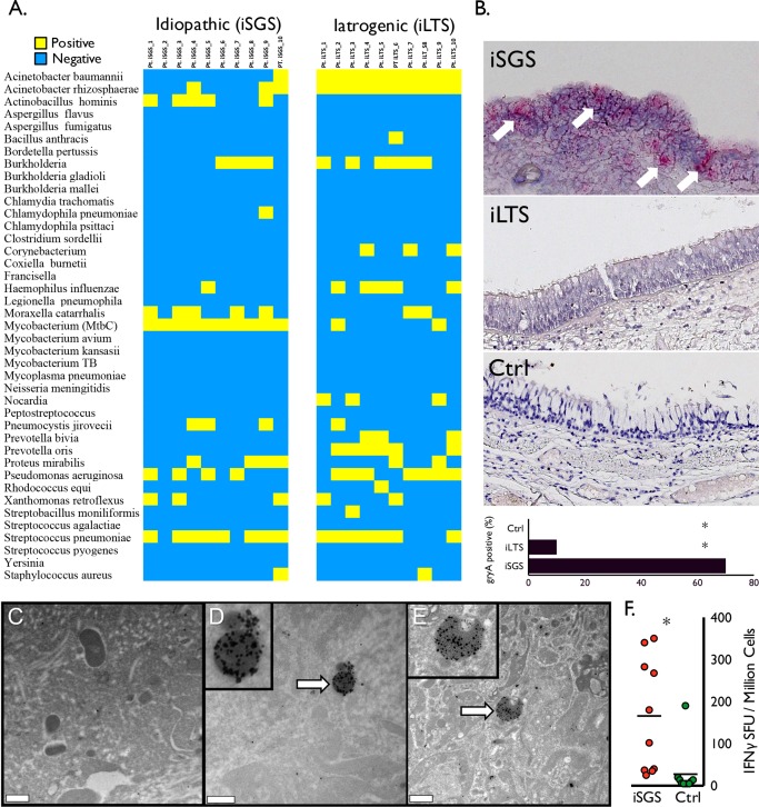
Figure 1.

Mycobacterium species in iSGS patients. The qPCR results for panel of respiratory pathogens from 10 iSGS and 10 iLTS patients. (A) Yellow indicates positive PCR products; blue indicates negative result. Ten of 10 iSGS patients had detectable PCR products for mycobacterium tuberculosis complex (MtbC), compared with two of 10 iLTS patients (two‐tailed, chi‐squared test with continuity correction; P < 0.001). (B) Representative images from in situ hybridization for RNA of Mycobacterium gene product gyraseA (arrows depicting positive signal in iSGS specimen). Accompanying summary graph depicting seven of 10 iSGS patients with detectable in situ hybridization signal, compared with one 10 iLTS and 0 of 10 controls (two‐tailed, chi‐squared test; asterisk denotes P < 0.001). Immunogold labeling with an anti‐MtbC antibody and high‐resolution transmission electron microscopy analyses revealed multiple structures with associated labels that exhibit typical size (500 nm–2 μm) and shape (coccoid or bacillus) of Mycobacterium spp. Treatment with secondary antibody alone (not shown) or an unrelated antibody to Haemophilus influenzae (C) revealed sparse labeling that was significantly less than the labeling achieved with the anti‐Mtb treatment (D, E). Distribution of IFN‐γ production from ESAT‐6 stimulated peripheral blood mononuclear cells isolated from the peripheral blood of iSGS patients (red; n = 10) or healthy controls (green; n = 10). Bars represent the median (50th percentile), asterisk denotes significance (two‐tailed, Mann Whitney test; P < 0.005) (F). Ctrl = control; IFN‐γ = interferon gamma; iLTS = iatrogenic laryngotracheal stenosis; iSGS = idiopathic subglottic stenosis; MtbC = mycobacterium tuberculosis complex; PCR = polymerase chain reaction; qPCR = quantitative polymerase chain reaction.
