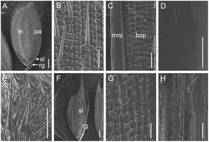
FIGURE 2.
scanning electron microscopy (SEM) analysis of glumes in the wild-type and the m34-z mutant at heading stage. (A–E) Wild-type spikelet. (B) Epidermal surface of the wild-type lemma. (C) Epidermal surface of the wild-type palea. (D) Epidermal surface of the wild-type sterile lemma. (E) Epidermal surface of the wild-type rudimentary glume. (F–H) m34-z mutant spikelet. (G) Epidermal surface of the m34-z mutant sterile lemma. (H) Epidermal surface of the m34-z mutant rudimentary glume. rg, rudimentary glume; sl, sterile lemma; le, lemma; pa, palea; bop, body of palea; mrp, marginal region of palea. Bars = 1000 μm in (A,F) and 100 μm in (B–E,G–H).

