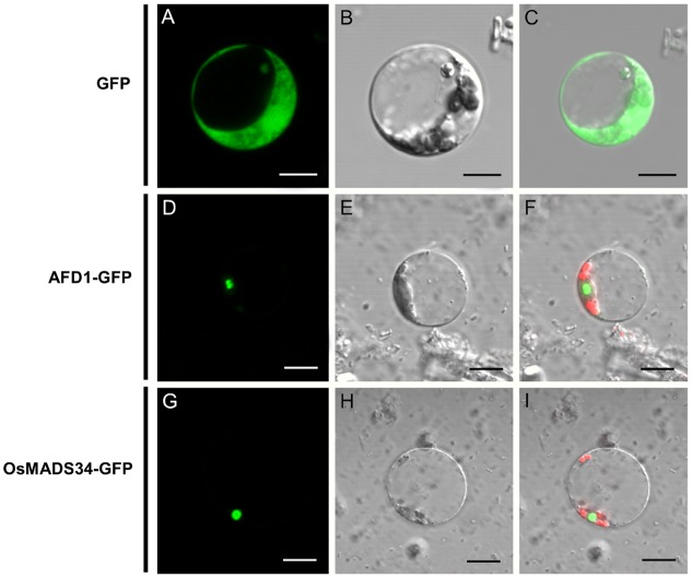
FIGURE 8.
Subcellular localization of the OsMADS34 protein. (A–C) GFP fusion protein. (A) differential interference contrast (DIC) image; (B) bright-field image; (C) merged image of GFP fusion protein. (D–F) AFD1-GFP. (D) DIC image; (E) bright-field image; (F) merged image of AFD1-GFP fusion protein. (G–I) OsMADS34-GFP. (G) DIC image; (H) bright-field image; (I) merged image of OsMADS34-GFP fusion protein. Bars = 5 μm in (B–G).

