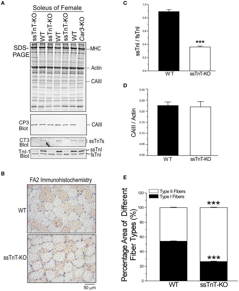
Figure 11.
Expression of CAIII in soleus muscle of a mouse model of Tnnt1 myopathy. (A) The expression level of CAIII was examined in adult soleus muscle of WT and Tnnt1-KO (ssTnT-KO) mice. The Western blot and glycerol-SDS-gel demonstrate the loss of slow TnT in Tnnt1-KO soleus muscle and the switches to significantly higher contents of fast types of myofilament proteins (the decreases in ssTnI/fsTnI ratio). Despite the diminished slow fiber content, the level of CAIII expression was remained. (B) Immunohistochemistry staining using anti-MHC I mAb FA2 of cross sections of Tnnt1-KO soleus muscle showed atrophy of type I slow fibers and hypertrophy of type II fast fibers as compared with WT control. (C) Densitometry quantification of the TnI-1 mAb Western blots confirmed the significantly decreased ratio of ssTnI/fsTnI in Tnnt1-KO soleus muscle in comparison with that of WT soleus muscle. (D) Densitometry quantification of Western blot normalized to the actin band in SDS-gel confirmed no decrease of CAIII in Tnnt1-KO soleus muscle vs. the WT control. (E) Quantification of the percentage cross sectional areas of type I and type II fibers in soleus muscles confirmed the atrophy of type I fibers and hypertrophy of type II fibers in Tnnt1-KO mice vs. that in WT mice. The data are shown as mean ± SE. N = 3 mice each in WT and Tnnt1-KO groups. ***P < 0.001 as compared with WT using Student's t-test.

