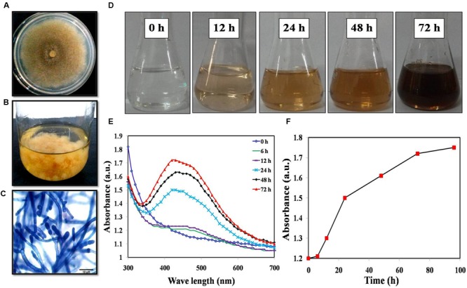
FIGURE 1.

(A) Colony morphology; (B) Mass culture of Mucor hiemalis; (C) Microscopic view of M. hiemalis processed with lactophenol cotton blue stain; (D) Optical images of biosynthesized AgNP showing a range of colors from colorless (start of biosynthesis) to reddish-brown after 24 h; (E) UV-Vis spectra of M. hiemalis-derived AgNPs at various reaction times; (F) Reaction saturation curve indicating the evolution of the absorption band as a function of time.
