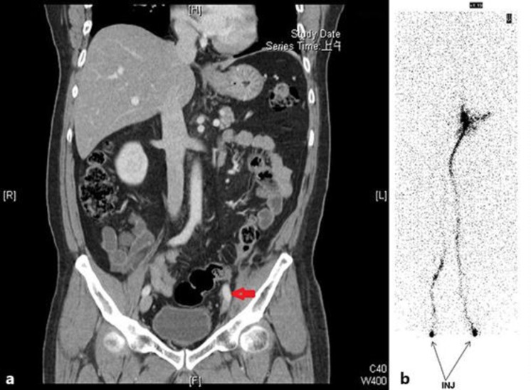
Fig. 3.
A 51-year-old man had left ureteral urothelial carcinoma with mesenteric seeding. He presented with left lower-extremity swelling. The CT scan revealed tumor compression of the left iliac vein with narrowing (coronary view) (a). Radionuclide venography showed a left iliac vein obstruction (b).

