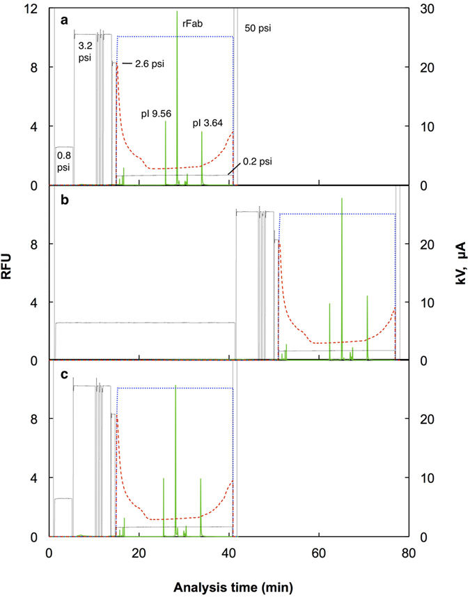
Figure 4. Analysis of rFab using the unified capillary.

The rFab was applied to the unified capillary consisting of anti-E tag antibody column (50 μm i.d.; 18.5 cm long) and PDMA-coated capillary (50 μm i.d.; 30 cm long). After sample application at 0.8 psi, the capillary was rinsed and filled with the carrier ampholyte solution containing 2.5 nM each of pI 9.56 and 3.64 markers at 3.2 psi. The anode solution, 100 mM phosphoric acid, was then injected to fill the antibody column segment of the capillary at 2.6 psi for 1 min and IEF was started by applying +25 kV and 0.2 psi at the antibody column side, and with 100 mM NaOH as the cathode solution. The focused protein and markers were detected with a fluorescence detector placed at 10 cm from the cathodic end. (a) 50 nM of labeled rFab in PBS-Tw, 4 min injection, 24 fmol; (b) 5 nM of labeled rFab in PBS-Tw, 40 min injection, 24 fmol; (c) 50 nM of labeled rFab in 50% serum, 4 min injection, 24 fmol. Green solid lines, fluorescence; blue dotted lines, voltage; red broken lines, current; black lines, pressure.
