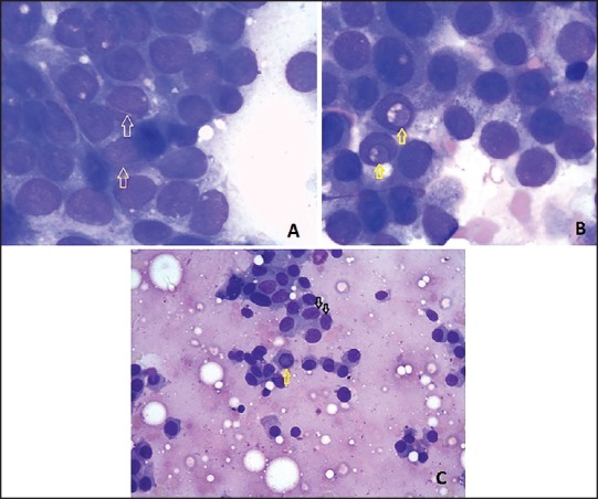
Figure 2.

Cytology of cases reported as “suspicious of papillary carcinoma”: (a) Showing nuclear grooves (arrows). This case on histopathology was found to be of adenomatous goiter. (MGG stain, ×100) (b) Showing intranuclear cytoplasmic inclusions (arrows). This case on histopathology was found to be nodular goiter. (MGG stain, ×100) (c) Showing nuclear grooves (black arrows) and intranuclear cytoplasmic inclusions (yellow arrow). This case on histopathology was found to be Hashimoto's thyroiditis. (MGG stain, ×40)
