Figure 1.

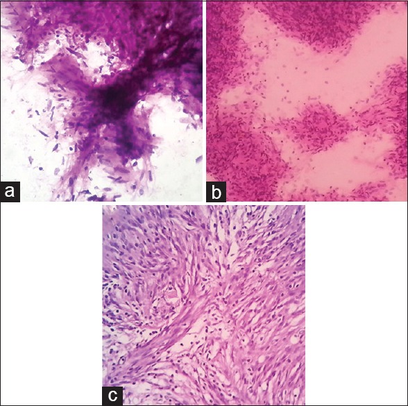
(a) Central nervous system (CNS) squash cytology of schwannoma (H&E stain, ×400); (b) CNS squash cytology [showing whorls of meningioma] of meningioma (H&E stain, ×400); (c) Histopathology of meningioma (H&E stain, ×400)

(a) Central nervous system (CNS) squash cytology of schwannoma (H&E stain, ×400); (b) CNS squash cytology [showing whorls of meningioma] of meningioma (H&E stain, ×400); (c) Histopathology of meningioma (H&E stain, ×400)