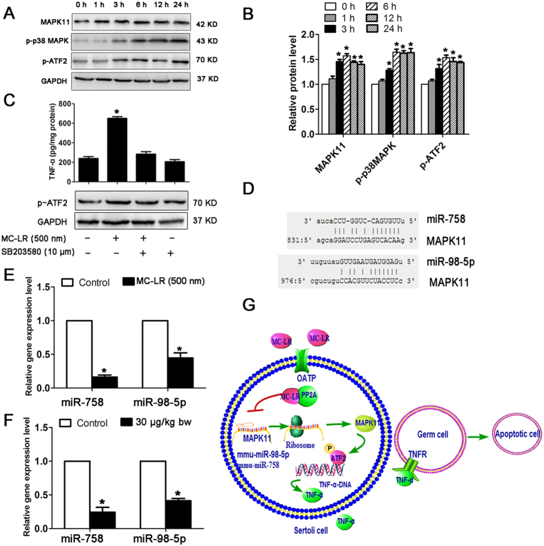
Figure 10. miR-98-5p and miR-758 regulatory networks involved in microcystin-leucine arginine (MC-LR)-induced tumor necrosis factor-α (TNF-α) production in Sertoli cells.
(A) Protein levels of mitogen-activated protein kinase 11 (MAPK11, p38 β isoform), p-p38 MAPK, and p-ATF2 in Sertoli cells cultured with medium supplemented with 500 nm MC-LR for various durations were examined by western blotting. (B) Expression levels of the proteins were quantified by densitometry and normalized to the expression of GADH. Densitometry data are shown as mean ± SD. *P < 0.05, compared with the control. (C) TNF-α production in Sertoli cells treated with MC-LR or SB203580. Cells were pre-incubated with 10 μm SB203580 for 1 h, followed by another incubation with 500 nm MC-LR for 24 h. TNF-α levels in cells were measured with ELISA kits (mean ± SD. *P < 0.05, compared with the control). (D) Predicted binding sites of miR-98-5p and miR-758 in the MAPK11 3′-UTR region are indicated. (E,F) The expression levels of miR-758 and miR-98-5p in Sertoli cells (E) and testes (F) were measured by q-PCR, and relative mRNA abundance was calibrated based on the miRNA amount of the corresponding gene in the untreated group (mean ± SD. *P < 0.05, compared with the control). (G) A model for the regulation of TNF-α gene expression by miRNAs is proposed. MC-LR stimulation significantly inhibits miR-98-5p and miR-758 expression, resulting in enhanced expression of MAPK11. MAPK11 is activated by p38 signaling cascade and subsequently phosphorylates transcription factor ATF-2. Phosphorylated ATF-2 binds to the promoter of TNF-α to stimulate TNF-α expression. Secreted TNF-α can bind to the tumor necrosis factor receptor 1 (TNFR1) of the surrounding cells and induce cell apoptosis.

