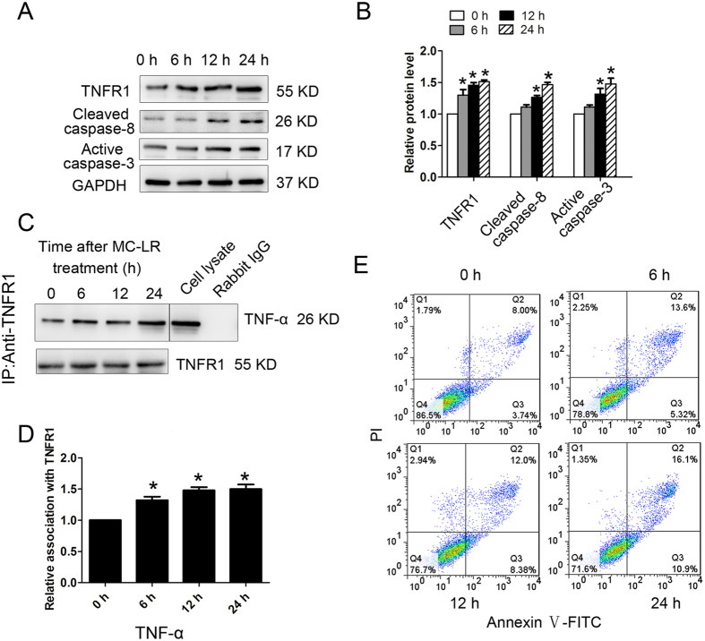
Figure 11. Tumor necrosis factor-α (TNF-α)-induced germ cell apoptosis via interacting with tumor necrosis factor receptor (TNFR1).
Germ cells were treated with 20 ng/mL TNF-α for various durations (0 h, 6 h, 12 h, and 24 h). (A,B) The expression of TNFR1, cleaved caspase-8, and active caspase-3 in germ cells at specified time points were examined by western blotting. Representative gel electrophoresis bands are shown (A), and the expression levels of the proteins were quantified by densitometry and normalized to the expression of GAPDH (B). Densitometry data are shown as mean ± SD. *P < 0.05, compared with the control. (C) Germ cell lysates were subjected to coimmunoprecipitation (Co-IP) with anti-TNFR1 antibody, and the blot was probed with anti-TNF-α antibody. Moreover, blots were re-probed with anti-TNFR1 antibody to confirm equal protein loading. Co-IP with rabbit IgG served as a negative control. The presence of TNF-α in the cell lysate was detected by western blotting, serving as a positive control. (D) Densitometric analysis of data shown in C is plotted, and the relative association at time 0 h was arbitrarily set as 1. Data are shown as mean ± SD. *P < 0.05, compared with the control. (E) The number of apoptotic cells was determined by flow cytometric analysis. Cells were stained with FITC-conjugated annexin V and PI and then were analyzed by flow cytometry.

