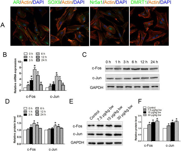
Figure 2. Induction of proto-oncogene expression in Sertoli cells and testes by microcystin-leucine arginine (MC-LR).
(A) Mouse Sertoli cells were collected and cultured for 2 days. Representative immunofluorescence analyses of the marker proteins of Sertoli cells are shown (×600). Nuclei were stained with DAPI (blue), and phalloidin (orange) was used to label the actin. Sertoli cells were treated with 500 nm MC-LR for various durations as indicated. (B) Changes of c-Fos and c-Jun transcript were examined by q-PCR, and relative mRNA abundance was calibrated based on the mRNA amount of the corresponding gene in the untreated group (mean ± SD. *P < 0.05, compared with the control). (C,D) Expression of c-Fos and c-Jun in Sertoli cells was detected by western blotting. Representative gel electrophoresis bands are shown (C), and the expression levels of the proteins were quantified by densitometry and normalized to the expression of GAPDH (D). Densitometry data are shown as mean ± SD. *P < 0.05, compared with the control. Male mice were injected intraperitoneally with saline or MC-LR (7.5 μg/kg body weight (bw), 15 μg/kg bw, and 30 μg/kg bw) for 7 days. (E,F) The expression levels of c-Fos and c-Jun were analyzed by western blotting. Representative gel electrophoresis bands are shown (E), and the expression levels of the proteins were quantified by densitometry and normalized to the expression of GAPDH (F). Densitometry data are shown as mean ± SD. *P < 0.05, compared with the control.

