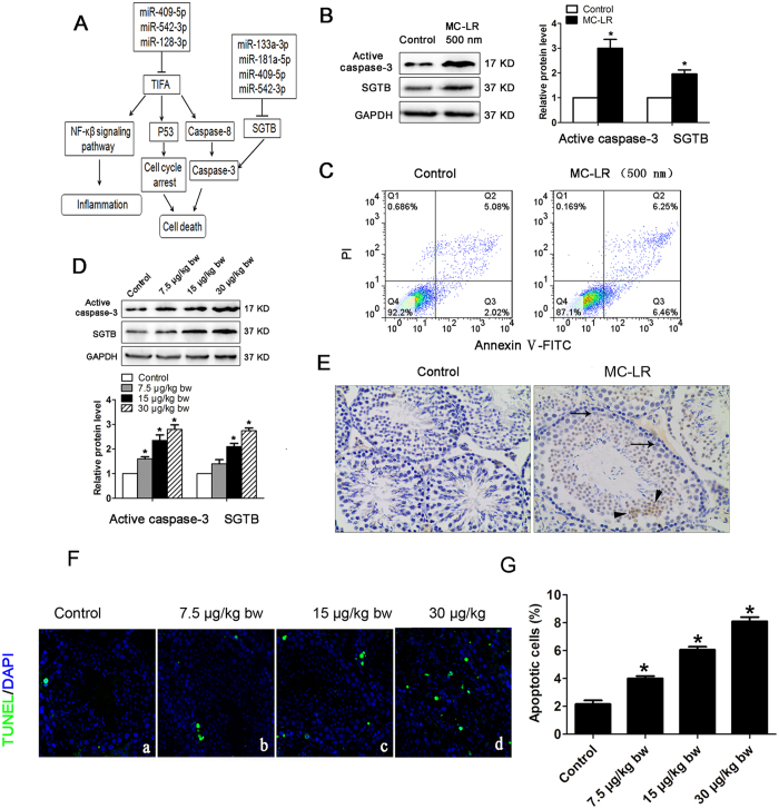
Figure 5. Induction of cell apoptosis by Microcystin-leucine arginine (MC-LR).
(A) A putative model showing TNF receptor associated factor (TRAF)-interacting protein with a forkhead-associated (FHA) domain (TIFA) and small glutamine-rich tetratricopeptide repeat (TPR)-containing β (SGTB) are involved in MC-LR-induced cell apoptosis. (B) The protein levels of SGTB and active caspase-3 in Sertoli cells treated with MC-LR (500 nm) were measured by western blotting, and the expression levels of the proteins were quantified by densitometry and normalized to the expression of GAPDH. Densitometry data are shown as mean ± SD. *P < 0.05, compared with the control. (C) The number of apoptotic cells was determined by flow cytometric analysis. Cells were stained with FITC-conjugated annexin V and PI and then were analyzed by flow cytometry. (D) The protein levels of SGTB and caspase-3 in mouse testes were examined by western blotting, and the expression levels of the proteins were quantified by densitometry and normalized to the expression of GAPDH. Densitometry data are shown as mean ± SD. *P < 0.05, compared with the control. (E) Expression of active caspase-3 was examined by immunohistochemistry (×200). Arrows and arrowhead indicate Sertoli cells and germ cells. (F) Apoptotic cells of testicular tissues were determined by terminal deoxynucleotidyl transferase-mediated dUTP nick end labeling (TUNEL) assay (×600). Nuclei were stained with DAPI (blue) and green dots indicate apoptotic cells. (a) Control, (b) 7.5 μg/kg body weight (bw), (c) 15 μg/kg bw, (d) 30 μg/kg bw. (G) Apoptotic index was calculated as the percentage of TUNEL positive cells in all cells in an image (mean ± SD. *P < 0.05, compared with the control).

