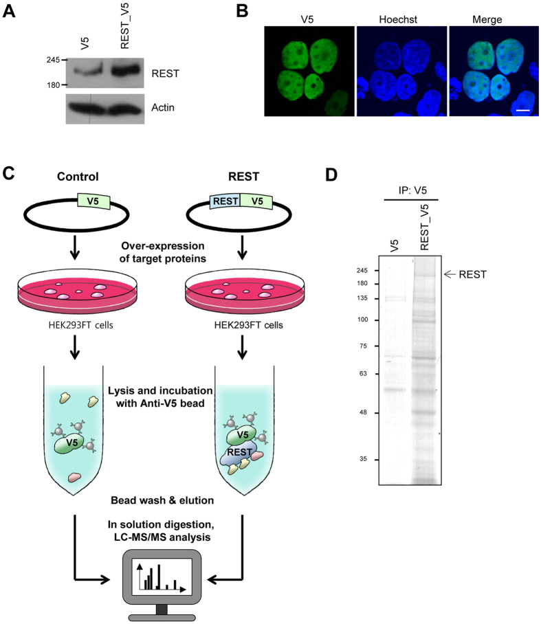
Figure 1. Experimental scheme and analysis of REST-interacting proteins.
(A) Western blot analysis of REST and actin. HEK293FT cells were transfected with control and REST-V5 expressing vector, respectively. Levels of REST and actin were assessed by western blotting. (B) Immunofluorescence staining to confirm localization of transiently expressed REST. HEK293FT cells transfected with the REST-V5 expressing vector were fixed after 24 h, followed by probing with V5 antibody. Images analyzed at magnification of 400x. Scale bar indicates 10 μm. (C) Experimental procedures for LC-MS/MS analysis after immunoprecipitation of REST. V5-tagged REST proteins were expressed in HEK293FT cells and immunoisolated using anti-V5 antibody immobilized on agarose beads. Immunocomplexes were eluted and digested for LC-MS/MS analysis. (D) SDS-PAGE and Coomassie blue staining of immunocomplexes. The arrow indicates REST. Full-length gels and blots are included in the Supplementary Figure 3.

