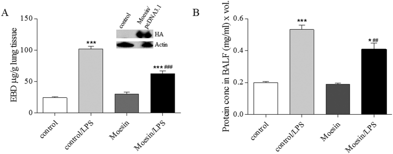
Figure 7. Moesin phosphorylation plays an essential role in LPS-induced lung injury in vivo.
PcDNA3.1 plasmids (40 μg) encoding empty vector (control) or unphosphorylatable Moesin mutant were complexed with JetPEI transfection reagent. Three days later, LPS (1 mg/kg) was injected i.t. (A) Bar graph shows Evans Blue dye extravasation, reflecting capillary leak. Western blot of lung homogenates stained for HA-tagged Moesin-mutant. (B) Bar graph shows protein levels in BALF (an index of lung inflammation and capillary leak). *P < 0.05, ***P < 0.0001 vs. control, ##P < 0.01, ###P < 0.0001 vs. control/LPS.

