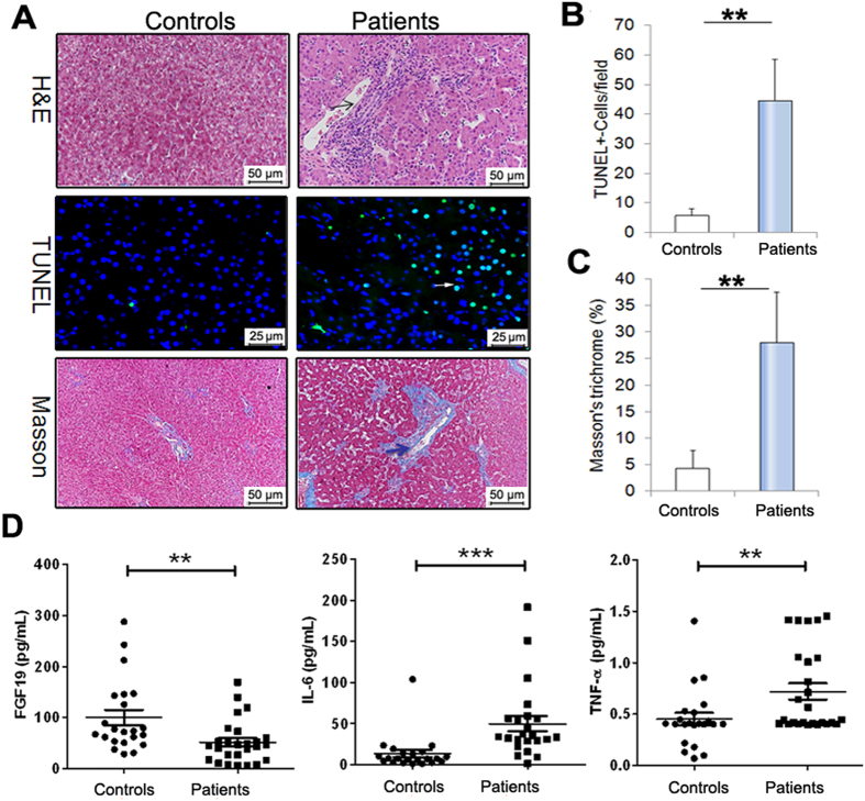
Figure 1. Liver histological changes in pediatric IF patients.
(A) Pediatric IF patients exhibit inflammatory infiltrate (H&E staining, n = 9), hepatic apoptosis (TUNEL-stained, n = 7) and fibrosis (Masson trichrome assay, n = 7). (B) Quantification of TUNEL-positive cells. (C) Quantification of Masson trichrome analysis. (D) Serum FGF19 concentration was significantly lower in pediatric IF patients compared to controls. Conversely, serum levels of IL-6 and TNF-α were significantly higher in patients than in controls. Scale bar = 25 or 50 μm. **p < 0.01; ***p < 0.001.

