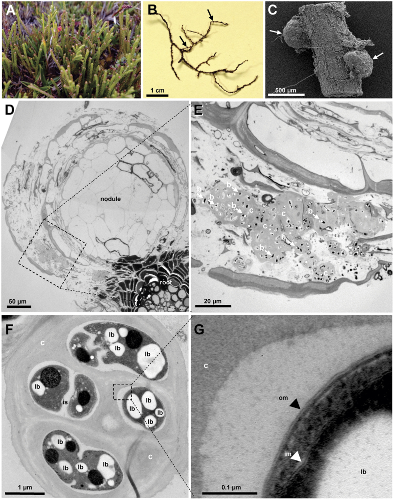
Figure 1.
Photograph (A), stereo microscope image (B), scanning electron (SEM; C) and transmission electron microscope (TEM; D–G) images of Lepidothamnus fonkii. Photograph of L. fonkii at the SKY field site at Seno Skyring (Southern Patagonia, Chile; A) and roots densely covered by nodules (B). Root nodules (arrows) were smaller than 500 μm in diameter (SEM; C). Ultrastructure of root with nodule (TEM; D) revealed capsules with multiple bacteria located primarily at the vicinity of the nodules (arrows indicate some of the bacterial cells; E) Enlarged capsules indicate ultrastructure of bacteria containing lipoid bodies (F). Intact outer and inner membranes (black and white arrowheads, respectively; G) of bacteria were indicative of living gram negatives, which is in agreement with active diazotrophic Beijerinckiaceae-related bacteria detected at the roots. Bar represents a scale bar (B–G). Squares and dashed lines indicate areas that were enlarged in the following panel. Abbreviations: b, bacteria; c, capsule; im, inner membrane; is, intercellular space; lb, lipoid bodies; om, outer membrane.

