
Figure 1. Accumulation of biotin in peripheral neural tissues.
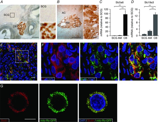
A and B, left: detection of biotin levels in slices of carotid artery bifurcation and adrenal gland. Scale bars 0.5 mm. Right: magnification of superior cervical ganglia (SCG), carotid body (CB) and adrenal medulla (AM) parenchyma indicated by the squares in the corresponding panels on left. Scale bars = 0.1 mm. C and D, Slc5a6 and SLC19a3 mRNA levels in CB and AM compared to SCG (mean ± SEM, n = 5 per group). **Statistical significance P < 0.01. E, fluorescence immunohistochemical detection of tyrosine hydroxylase (red) and endogenous biotin (green) in carotid body slices from control rats. Scale bar = 20 μm. F, magnification of the area inside the square in E to illustrate the co‐localization of biotin and TH. Scale bar = 10 μm. G, confocal image showing the immunofluorescent co‐localization of biotin with mito‐ro‐GFP expressed in a dispersed glomus cell by adenoviral infection. The mito‐ro‐GFP directs the expression of GFP to the mitochondrial matrix. Scale bar = 5 μm.