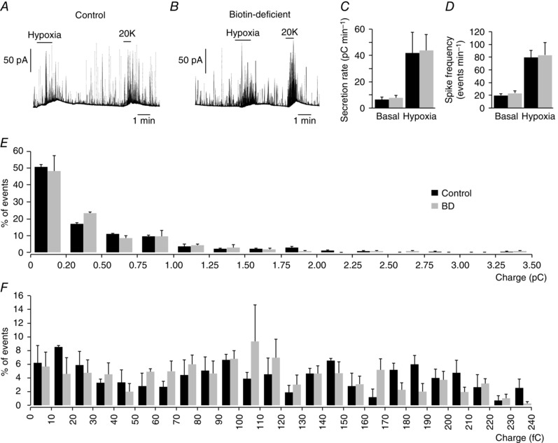
Figure 4. Secretory activity of chromaffin cells in adrenal medulla slices from control and biotin‐deficient animals.

A and B, amperometric recording of secretory responses of chromaffin cells to hypoxia and to depolarization with 20 mM K+ in control and biotin‐deficient animals. C, secretion rate in pC min−1 recorded under basal and hypoxic conditions from control (black, 41.45 ± 15.56 pC min−1, n = 5) and biotin‐deficient (grey, 43.74 ± 12.40 pC min−1, n = 5) animals. D, spike frequency obtained under basal and hypoxic stimulation of control (black, 80 ± 11 spikes min–1, n = 5) and biotin‐deficient (grey, 83 ± 19 pC min−1, n = 5) animals. E, frequency–charge distribution of individual exocytotic events recorded from chromaffin cells of control and biotin‐deficient rats in response to hypoxia (control, black, n = 311 spikes, 3 recordings; biotin‐deficient, grey, n = 445 spikes, 4 recordings). F, frequency–charge distribution of small vesicles (from 0 to 240 fC) recorded from chromaffin cells of control and biotin‐deficient rats (control, black, n = 152 spikes, 3 recordings; biotin‐deficient, grey, n = 221 spikes, 4 recordings). No statistically significant differences were observed in C–F.
