Fig. 3.

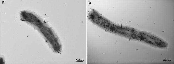
Transmission electron microscopy (TEM) micrographs of BCP1 cells grown for 120 h in the presence of 100 µg/mL (a), and 500 µg/mL (b) of K2TeO3. Arrows indicate the intracellular TeNRs produced by the BCP1 strain

Transmission electron microscopy (TEM) micrographs of BCP1 cells grown for 120 h in the presence of 100 µg/mL (a), and 500 µg/mL (b) of K2TeO3. Arrows indicate the intracellular TeNRs produced by the BCP1 strain