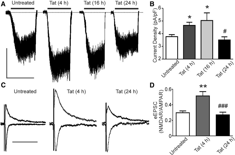
Figure 1.
HIV-1 Tat potentiates NMDAR-mediated currents that adapt to baseline during 24 h of exposure. A, Representative traces showing NMDA-evoked steady-state whole-cell currents from neurons voltage clamped at −60 mV after being treated for 0, 4, 16, or 24 h with Tat. NMDA (10 μm) was applied as indicated by the horizontal bars. Scale bar, 100 pA, 60 s. B, Bar graph summarizing the effects of 0, 4, 16, and 24 h treatment with Tat on the NMDA-evoked steady-state whole-cell current normalized to whole-cell capacitance. Data are mean ± SEM (n ≥ 4 for all groups). *p < 0.05 relative to untreated, #p < 0.05 relative to 4 and 16 h Tat-treated groups (one-way ANOVA with Tukey's post hoc test). C, Representative traces showing evoked EPSCs from neurons voltage clamped at −70 mV (AMPAR) and +40 mV (+30 ms taken as NMDAR EPSC) from cells treated for 0, 4, or 24 h with Tat. The presynaptic neuron was stimulated using a bipolar concentric electrode positioned near the soma. Traces were normalized to AMPAR currents. Scale bar, 100 ms. D, Bar graph summarizing the effects of Tat treatment for 0, 4, and 24 h on the NMDAR:AMPAR ratio of evoked EPSCs. Data are shown as mean ± SEM (n ≥ 6 for all groups). **p < 0.01 relative to untreated, ###p < 0.001 relative to Tat-treated (4 h) (one-way ANOVA with Tukey's post hoc test).

