Figure 5.
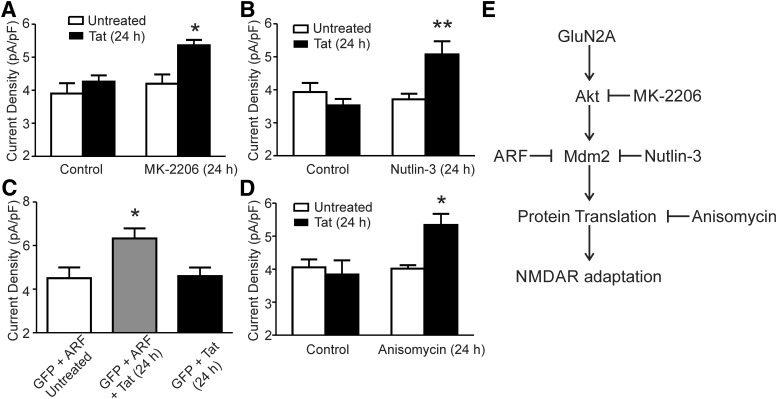
Inhibition of an Akt-Mdm2 signaling pathway prevents NMDAR adaptation to Tat. A, Bar graph summarizing the amplitude of NMDA-evoked (10 μm NMDA) steady-state currents normalized to whole-cell capacitance from neurons treated with or without Tat for 24 h in the presence or absence of the Akt inhibitor MK-2206 (1 μm). B, Bar graph summarizing the amplitude of NMDA-evoked steady-state currents normalized to whole-cell capacitance from neurons treated with or without Tat for 24 h in the presence or absence of the Mdm2 inhibitor Nutlin-3 (1 μm). C, Bar graph summarizing the amplitude of NMDA-evoked steady-state currents normalized to whole-cell capacitance from neurons transfected with GFP and ARF with or without Tat for 24 h and neurons transfected with GFP alone and treated with Tat for 24 h. D, Bar graph summarizing the amplitude of NMDA-evoked steady-state currents normalized to whole-cell capacitance from neurons treated with or without Tat for 24 h in the presence or absence of anisomycin (10 μm). Data are shown as mean ± SEM (n ≥ 6 for all groups). *p < 0.05, **p < 0.01 relative to all other groups (one-way ANOVA with Tukey's post hoc test). E, Scheme showing the adaptation pathway activated during 24 h of exposure to HIV Tat. Signaling elements were identified based on inhibition by the treatments shown.

