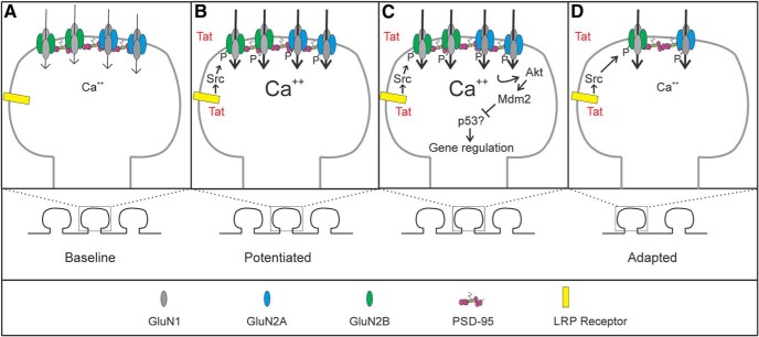
Figure 7.
Schematic of signaling events involved in Tat-induced changes in NMDARs. Schematic shows the hypothesized changes in NMDAR function during exposure to HIV-1 Tat. At baseline (A), NMDAR-mediated Ca2+ signaling and synaptic number are at homeostasis. After exposure to Tat (B), Tat is internalized via the LRP receptor and induces potentiation of Ca2+-influx through NMDARs via a Src-dependent phosphorylation of both GluN2A and GluN2B-containing NMDARs. Prolonged potentiation (C) of GluN2A-containing NMDARs leads to an adapted state via an Akt-Mdm2 signaling pathway that is dependent on protein translation. The adapted state (D) contains fewer synapses and Ca2+ influx has adapted in remaining spines.

