Figure 2.
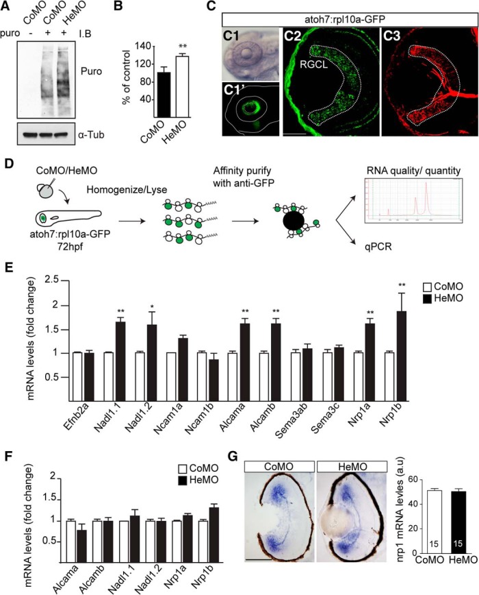
Hermes exerts a negative translational control of specific mRNAs in RGCs. Increase in puromycin incorporation detected by Western blotting in the Hermes-depleted condition compared with control (A, B). GFP expression is restricted to the eye in the atoh7:rpl10a-GFP transgenic line (C1, C1′), with a positive signal in Zn-5 positive RGCs and photoreceptors (C2, C3). atoh7:rpl10a-GFP embryos at 72 hpf, injected with CoMO or HeMO, were homogenized and immunoprecipitations against GFP were performed on lysates. The total RNA was then extracted from the ribosome–mRNA complexes, analyzed by bioanalyzer, and quantified by quantitative RT-PCR (D). Quantifications show an increase of nadl1.1, nadl1.2, alcama, alcamb, nrp1a, and nrp1b mRNAs bound to ribosomes in absence of Hermes (E). Quantifications of total RNAs input show no difference between CoMO and HeMO conditions (F). nrp1 in situ hybridization on 72 hpf retinal transverse sections shows no difference between HeMO- and CoMO-injected embryos (G). Quantifications of signal intensity show no difference in nrp1 expression in HeMO compared with CoMO (G). mRNA levels were calculated by using the formula 2−ΔΔCt with β-actin mRNA as a calibrator (E, F). Error bars indicate SEM. Numbers of embryos analyzed are indicated on bars. Scale bars: C2, C3, 30 μm; G, 100 μm.

