Figure 4.
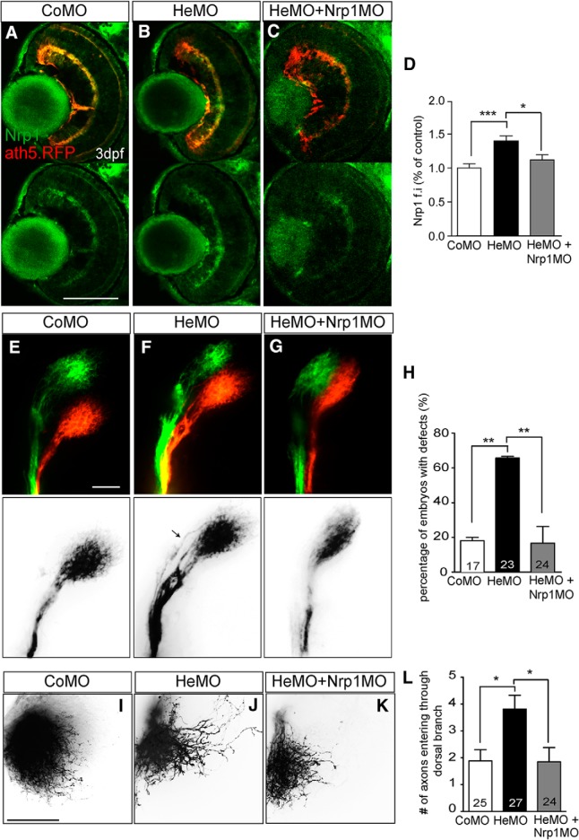
Restoring Nrp1 levels rescues the dorsal axon topography defect in Hermes-depleted embryos. Shown is Nrp1 immunostaining on retina transverse sections from atoh7:gapRFP zebrafish embryos injected with CoMO (A), HeMO (B), or HeMO+Nrp1MO (C). Quantifications show a significant increase of Nrp1 signal intensity in the HeMO condition compared with CoMO and HeMO + Nrp1MO (D). Lateral view of whole-mount DiI- and DiO-injected retina from CoMO- (E), HeMO- (F), and HeMO + Nrp1MO (G)-injected embryos. HeMO + Nrp1MO-coinjection showed a significant reduction of the percentage of embryos with mistargeting in the OT compared with HeMO (H). DiI-labelling of dorsal axons in the tectum of CoMO (I), HeMO (J), and HeMO + nrp1MO (K) injected embryos. Quantifications show a rescue of misprojecting dorsal axons in HeMO+nrp1MO injected embryos (L). Error bars indicate SEM. Numbers of embryos analyzed are indicated on bars. Scale bars: A–C, 100 μm; E–G, I–K, 50 μm).

