
Figure 4. CB1R agonists do not evoke Ca2+ transients via the IP3 pathway in myotubes and adult muscle fibres.
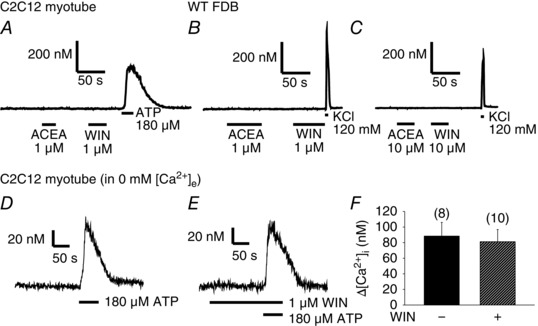
Representative traces of single‐cell [Ca2+]i measurements performed on Fura‐2‐loaded (A) C2C12 myotubes (n = 51) and (B) FDB fibres of WT mice (n = 53). The CB1R agonists 1 μm ACEA and 1 μm WIN did not evoke Ca2+ transients as opposed to 120 mm KCl or 180 μm ATP used as viability controls. Even when applied at 10 μm (C), these agonists could not evoke Ca2+ transients as opposed to KCl in FDB fibres (n = 13). However, the IP3 pathway was clearly activated in C2C12 myotubes by the administration of 180 μm ATP in Ca2+‐free Tyrode solution in the (D) absence and (E) presence of 1 μm WIN. F, pooled data of the amplitude of the ATP‐evoked transients.