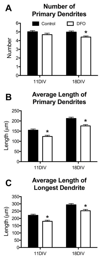
Figure 5. Iron deficiency impairs primary dendrite development in cultured hippocampal neurons.

Hippocampal neurons cultured from E16 mice were transfected with an hrGFP-expressing plasmid and then treated with DFO and 5-FU beginning at 3DIV. At 11DIV and 18DIV cultures were fixed and processed for hrGFP and MAP2 immunocytochemistry. Images were taken at 20x magnification and merged to allow manual tracing of dendrites on individual neurons. The (A) number and (B) average length of primary dendrites were quantified. (C) As an in vitro surrogate for apical dendrite length, the average length of the longest dendrite was quantified. The data from 2–3 independent cultures were pooled and are presented as mean ± SEM. Asterisks indicate statistical difference between groups at a given neuronal age by Student’s t-test (p < 0.05). 11DIV: n=72–84 neurons per group, 18DIV: n=83–90 neurons per group.
