
Fig. 4.
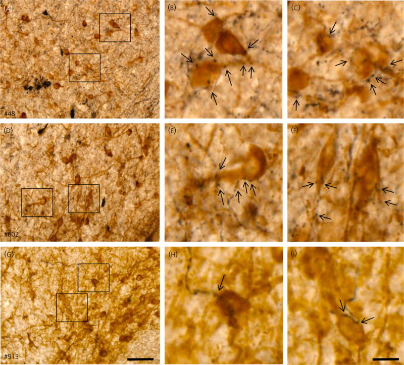
Arcuate nucleus (ARC) projections of retrochiasmatic area neurones in three ewes. Demonstration of biotinylated dextran amine (BDA)-labelled axons in ARC in close proximity to kisspeptin immunoreactive cell bodies in ewes #48 (A–C), #602 (D–F), and #914 (G–I). (A, D, G) show low magnification overview of labelling in ARC; scale bar = 50 μm. (B, C, E, F, H, I) show higher magnifications of areas indicated by boxes in (A, D, G) and illustrate BDA-labelled boutons in close proximity to kisspeptin neurones (indicated by arrows); scale bar = 15 μm.