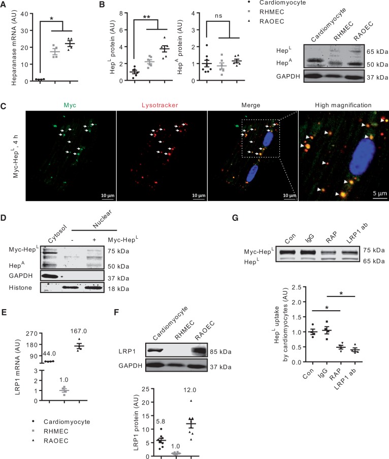
Figure 3.
Cardiomyocytes are also capable of HepL uptake. Cell lysates of primary rat cardiomyocytes, RAOEC or RHMEC were obtained for determination of heparanase mRNA (A) and protein (B), n = 4–8. Cardiomyocytes seeded on coverslips were placed in a 6-well plate, and treated with 500 ng/mL myc-HepL prior to immunofluorescence staining examined under a confocal microscope. The merged image of heparanase and lysosomes is described in the third (scale bar, 10 µm) and fourth (scale bar, 5 µm) panels from left (C) and are data from a representative experiment. Isolated myocytes were also treated with or without myc-HepL for 4 h. Following this incubation, nuclear and cytosolic fractions were isolated, and HepA protein levels determined by western blot (D). Cell lysates of primary rat cardiomyocytes, RAOEC, or RHMEC were obtained for determination of LRP1 mRNA (E) and protein (F), n = 4 and n = 8. In a different experiment, in cardiomyocytes incubated with HG, cells were pre-treated with or without 400 nM RAP or 40 μg/mL LRP1 neutralizing antibody for 1 h prior to incubation with 500 ng/mL myc-HepL for 4 h. Cell lysates were collected to determine HepL uptake, n = 4 (G). 40 μg/mL IgG was used as a control for the LRP1 neutralizing antibody experiment. *P < 0.05, **P < 0.01.

