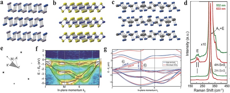
Figure 1.

a–c) Crystal structures of layered orthorhombic, hexagonal, monoclinic phases of 2D GIVMCs. d) Raman spectra of bulk SnS2 at two excitation wavelengths (532, 633 nm). Gray and black vertical lines mark the main Raman lines of 2H–SnS2 and 4H–SnS2. e) Low‐energy electron diffraction pattern with overlaid irreducible wedge of the surface Brillouin zone. f) Angle resolved photoelectron spectroscopy of the projected band structure of bulk SnS2. False color scale: Dark blue, lowest intensity; white, highest intensity. Black lines are results of DFT band structure calculations for 4H–SnS2 using the HSE hybrid functional. g) Comparison of the calculated band structure of bulk (4H) and monolayer SnS2. (d–g) Reproduced with permission.72 2014, American Chemical Society.
