
Figure 6.
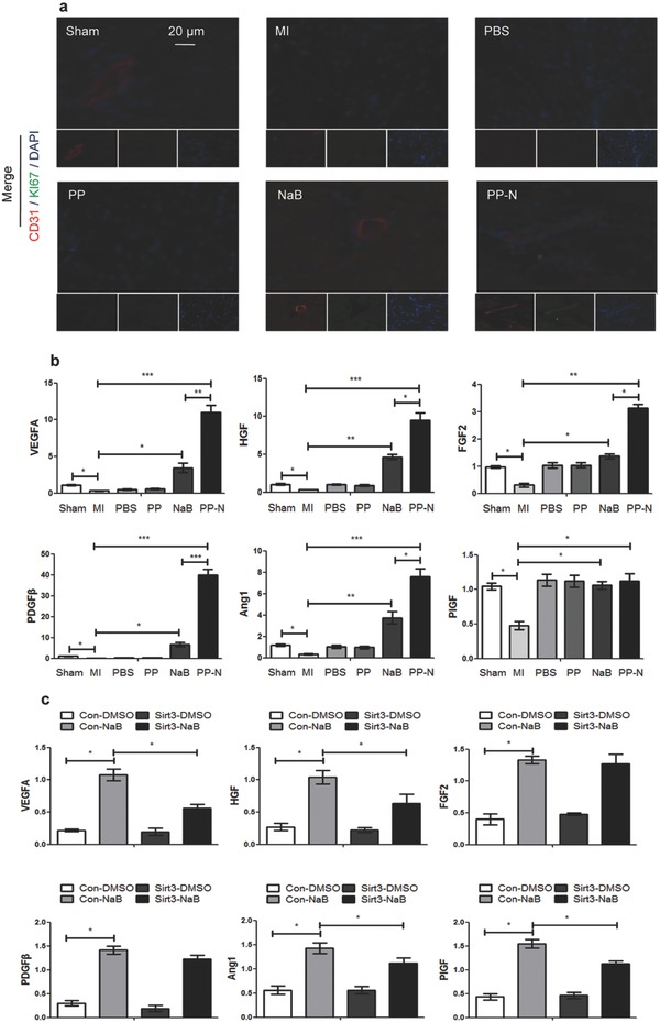
The effect of PP‐N on angiogenesis with Sirt3 activation. a) The heart sections collected at four weeks after injection were analyzed with immunofluorescence. They were stained for CD31 (red), a marker of endothelial cells, Ki67 (green), a marker of proliferation, and DAPI (blue), a maker of nuclei. b) qRT‐PCR analysis of the relative mRNA levels of angiogenesis‐related genes in the cardiac ischemic zone at four weeks after injection. c) RCMs were infected with pSUPER vectors expressing Sirt3 siRNA or with control vectors, as described in the protocol, and the cells were then treated with 200 × 10–6 m NaB for 24 h. The cytokines levels secreted by the RCMs were analyzed by enzyme‐linked immunosorbent assay (ELISA). *P < 0.05; **P < 0.01; ***P < 0.005.