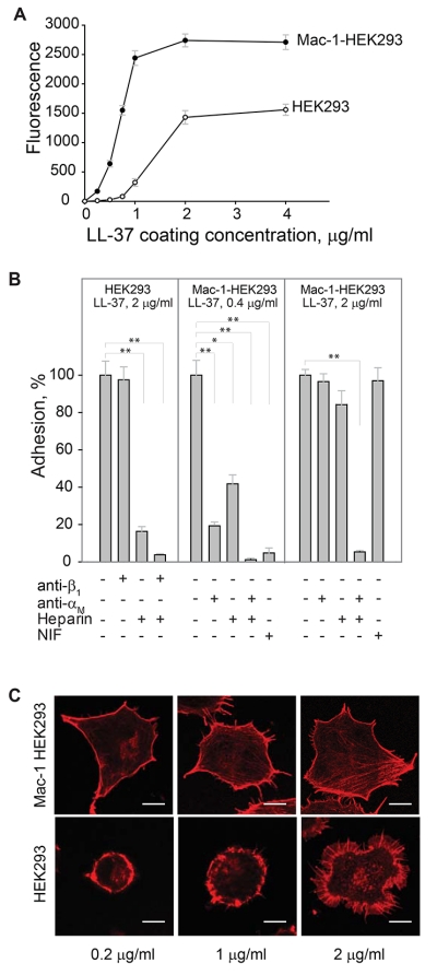
Figure 2. LL-37 supports adhesion of the αMβ2-expresing cells.
(A) Aliquots (100 μl; 5×10⁴/ml) of Mac-1-expressing HEK293 (Mac-1-HEK293) and wild-type HEK293 (HEK293) cells labeled with calcein were added to microtiter wells coated with different concentrations of LL-37 and postcoated with 1% PVP. After 30 min at 37 °C, nonadherent cells were removed by washing and fluorescence of adherent cells was measured in a fluorescence reader. A representative of 6 experiments in which two cell lines were tested side by side is shown. Data presented are means for triplicate determinations, and error bars represent S.E. (B) Wild-type HEK293 cells were preincubated with anti-β1 mAb (10 μg/ml), heparin (20 μg/ml) or their mixture for 15 min at 22 °C and added to wells coated with 2 μg/ml LL-37. Mac-1-HEK293 cells were preincubated with anti-αM mAb 44a (10 μg/ml), heparin (20 μg/ml) of their mixture and added to wells coated with 0.4 or 2 μg/ml LL-37. Adhesion was quantified as described above. Adhesion in the absence of Mac-1 inhibitors and heparin was assigned a value of 100%. Data shown are means ± S.E from 3-4 separate experiments with triplicate measurements. **p≤0.01, *p≤0.05 compared with control adhesion in the absence of inhibitors. (C) Mac-1-HEK293 (upper panel) and HEK293 cells (bottom panel) were plated on glass slides and allowed to adhere for 30 min at 37 °C. Nonadherent cells were removed and adherent cells were fixed with 2% paraformaldehyde followed by staining with Alexa Fluor 546 phalloidin. The cells were imaged with a Leica SP5 laser scanning confocal microscope with a 63× objective.

