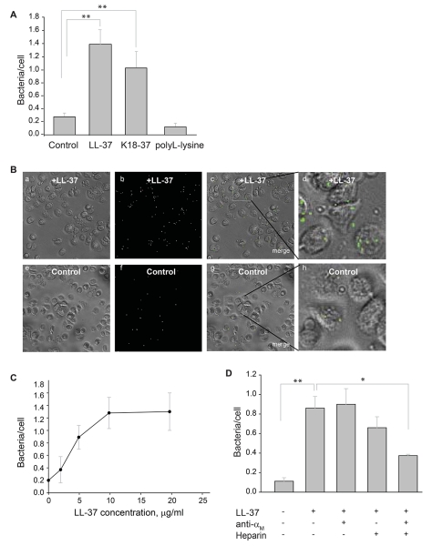
Figure 6. Effect of LL-37 on phagocytosis of S. aureus particles and E. coli by suspensions of murine IC-21 macrophages.
(A) Fluorescent S. aureus particles (4×10⁷/ml) were preincubated with LL-37 (5 μg/ml), K18-37 (10 μg/ml) or polyL-lysine (10 μg/ml) for 20 min at 22 °C. Soluble peptide was removed by centrifugation. Peptide-coated particles were incubated with suspensions of IC-21 macrophages (106/ml) for 60 min at 37 °C and nonphagocytosed particles were separated from cells by filtering the suspensions using a 3-μm pore Transwell inserts. Macrophages were transferred to wells of 96-well plates and trypan blue was added to wells. The ratio of bacterial particles per macrophage was quantified for five random fields per well using a 20× objective. Data shown are mean/cell ± S.E. from five or more experiments. **p≤0.01, compared with untreated control S. aureus. (B) Bright field (a,e), fluorescence (b,f) and merged (c,g) images of IC-21 macrophages incubated with LL-37-coated (a-c) or uncoated (e-g) control bacteria. The representative low power (20×) fields are shown. Enlarged images (d,h) of macrophages shown in the boxed areas in c and g. Phagocytosed bacterial particles are labeled in green. (C) Concentration-dependent effect of LL-37 on phagocytosis of E. coli by macrophages. Fluorescently labeled E. coli cells were incubated with different concentrations of LL-37 for 20 min at 22 °C. After the removal of nonbound peptide by centrifugation, LL-37-coated E. coli were incubated with IC-21 macrophages (106/ml) for 60 min at 37 °C. Phagocytosis was determined as in Fig. 6A. Data are expressed as mean ratios of bacteria per macrophage ± SE. A representative of three experiments is shown. (D) Fluorescent S. aureus particles were preincubated with LL-37 (5 μg/ml) for 20 min at 22 °C. Soluble peptide was removed by centrifugation. Peptide-coated bacterial particles were incubated with IC-21 macrophages (106/ml) in the presence of anti-αM mAb M1/70 (20 μg/ml), heparin (20 μg/ml) or their mixture. After 60 min incubation at 37 °C, nonphagocytosed S. aureus particles were removed and phagocytosis was measured as described above. Data shown are mean bacterial particles/cell ± S.E. of five random fields determined for each condition and are representative of 3 separate experiments. **p≤0.01, *p≤0.05

