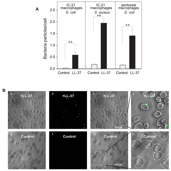
Figure 7. Augmentation of phagocytosis of LL-37-coated bacteria by adherent macrophages.
(A) Fluorescently labeled S. aureus particles and E. coli cells were incubated with LL-37 (5 μg/ml) for 20 min at 22 °C. Soluble peptide was removed by centrifugation and LL-37-coated bacteria were subsequently incubated with adherent IC-21 macrophages or mouse peritoneal macrophages for 60 min at 37 °C. Data are expressed as mean ratios of bacteria per macrophage ± SE. The panels shown in A are representative of three or more experiments. **p≤0.01 compared with untreated control bacteria. (B) A representative experiment showing bright field (a,e), fluorescence (b,f) and merged images (c,g) of IC-21 macrophages exposed to LL-37-coated and uncoated control S. aureus.

