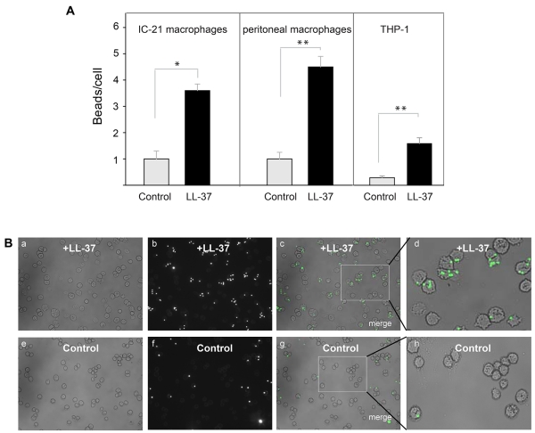
Figure 8. Effect of LL-37 on phagocytosis of latex beads.
(A) Fluorescent latex beads (2.5×10⁷/ml) were preincubated with LL-37 (10 μg/ml) for 20 min at 22 °C. Soluble peptide was removed from beads by high-speed centrifugation. Peptide-coated beads were incubated with IC-21 cells, mouse peritoneal macrophages or differentiated THP-1 cells for 30 min at 37 °C. Nonphagocytosed beads were separated from macrophages by centrifugation. The ratio of beads per macrophage was quantified from three fields of fluorescent images. Data shown are means ± S.E. of triplicate measurements and are representative of 3 experiments. **p≤0.01, *p≤0.05 compared with untreated beads. (B) Fluorescence of IC-21 macrophages exposed to LL-37-treated beads (a,b,c) or control untreated beads (e,f,g). Enlarged images of IC-21 macrophages showing phagocytosed LL-37-treated (d) or control (h) beads.

