Figure 1.

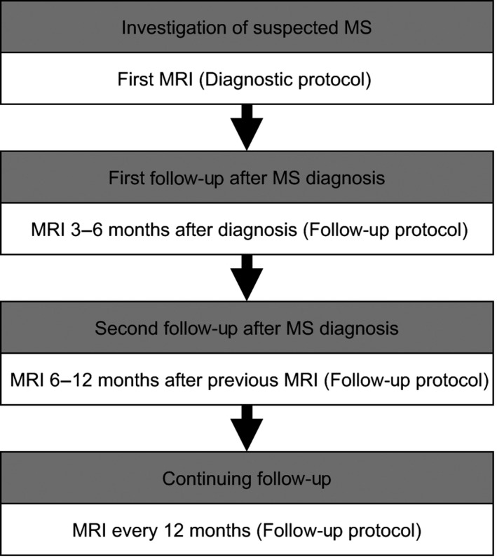
An example of timing of first, second, and continuing magnetic resonance imaging monitoring after multiple sclerosis diagnosis

An example of timing of first, second, and continuing magnetic resonance imaging monitoring after multiple sclerosis diagnosis