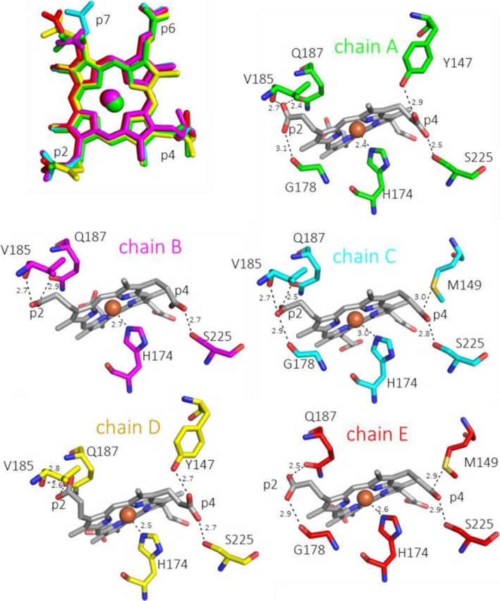
Figure 3.

Coproheme conformation and environment. Overlay of coproheme moieties bound to LmHemQ from all five subunits (top left), and H‐bonding interactions of coproheme propionates at positions 2 and 4 with the protein matrix in the respective subunits, residues within 3 Å of a propionate oxygen atom are depicted as sticks. The coordination of the proximal H174 to the coproheme iron is also shown.
