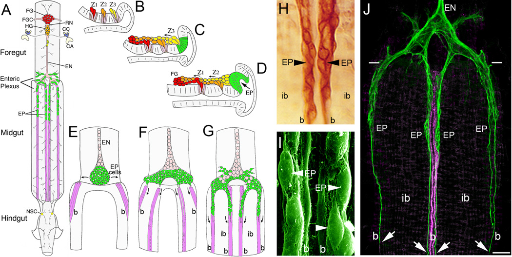
Figure 1. Embryonic development of the insect Enteric Nervous System (ENS) involves extensive patterns of neuronal and glial migration.
(A), Schematic drawing of the ENS and associated neurosecretory organs in the larval stage of the tobacco hornworm Manduca sexta (modified from [27]). The primary ganglion on the foregut is the frontal ganglion (FG; red), connected to the overlying brain lobes by paired frontal ganglion connectives (FGC). Several nerve branches extend anteriorly onto the pharynx, while the recurrent nerve (RN) extends posteriorly to the hypocerebral ganglion (HG; orange), situated below the brain. In Manduca, the hypocerebral ganglion initially forms during embryogenesis but then becomes closely opposed to the frontal ganglion and is no longer readily distinguished in later stages. The HG is also connected to the paired corpora cardiaca (CC; blue), the primary neurosecretory organs of the brain, which are adjacent to the corpora allata (CA; the source of Juvenile Hormone). From the HG, the esophageal nerve (EN) extends posteriorly along the length of the foregut, giving rise to short nerve branches that innervate the foregut musculature. Near the foregut-midgut boundary, the esophageal nerve connects with the enteric plexus that spans the foregut-midgut boundary, which includes nerve branches extending along radial muscles on the foregut and major nerves that extend along eight well-defined muscle bands that lie superficially on the midgut (purple). The enteric plexus contains a population of ~300 distributed neurons (EP cells; green), which includes intermingled groups of neurons expressing a variety of morphological and transmitter phenotypes. The EP cells occupy positions along the anterior 20% of the midgut, and extend long axons posteriorly along the muscle bands with sparse lateral branches that provide a diffuse innervation of the interband midgut musculature. The hindgut is innervated by branches of the proctodeal and rectal nerves that originate in the terminal abdominal ganglion of the ventral nerve cord. Branches of the proctodeal nerve also extend onto the posterior midgut and contain several peripheral neurosecretory cells (yellow). (B–C), Neurogenesis of the developing ENS in Manduca (after [31]). Panels show lateral views of the foregut midline; anterior is to the left, dorsal is to the top. When raised at 25°C, Manduca embryogenesis is complete in 100 hr (1 hr = 1 hour post-fertilization, or hpf). (B), By ~24 hpf, three neurogenic zones (Z1, Z2, & Z3) have formed in the dorsal foregut epithelium, which give rise to a series of mitotically active precursor cells via sequential delamination. Precursors giving rise to neurons typically divide only once (or occasionally twice) after delaminating, similar to midline precursors in the embryonic CNS. (C), By 28 hpf, streams of zone-derived cells have begun to migrate anteriorly along the foregut, while the remaining zone 3 cells delaminate as a group. The epithelium surrounding the original position of zone 3 subsequently differentiates into a distinct placode that will form the EP cells (green). (D), By 33 hpf, migrating zone cells have begun to form the frontal ganglion (FG), while the remaining zone 2 cells delaminate as a group. The EP cell placode has also begun to invaginate from the EP cell packet (described below). Zone 1 continues to generate cells until almost 40 hpf (not shown); late-emerging zone cells derived from all three zones tend to become glial precursors that remain mitotically active throughout much of embryogenesis and establish the glial sheath surrounding the foregut nerves and ganglia. (E–F), Formation of the midgut enteric plexus; panels show dorsal views of the developing ENS at the foregut-midgut boundary (after [88]). (E), By 40 hpf, the EP cells (green) have invaginated en mass from their neurogenic placode located within the posterior dorsal lip of the foregut (D, green). The neurons then commence a bilateral spreading phase of migration (arrows) that almost completely encircles the foregut, adjacent to the foregut-midgut boundary. Concurrently, subsets of longitudinal muscles on the midgut (magenta) begin to coalesce into eight well-defined bands as dorsal closure of the midgut proceeds. Anteriorly, the EP cell packet is in continuity with the developing esophageal nerve (EN), which contains populations of proliferating glial precursors (pink; derived from zone 3) that will subsequently ensheath the enteric plexus. (F), By 55 hpf, the EP cells have almost completely surrounded the foregut, and subsets of the neurons have aligned with each of the midgut muscle bands (only the dorsal four are shown). (G), By 58 hpf, subsets of EP cells have begun to migrate in a chain-like manner along the midgut muscle bands; smaller subsets also migrate onto radial muscles of the foregut (muscles not shown). Proliferating glial cells (pink) subsequently migrate along the pathways established by the neurons, thereby ensheathing the branches of the enteric plexus. (H), Magnified view of EP cell groups migrating on the mid-dorsal band pathways (at 58 hpf) of an embryo immunostained with an antibody recognizing all isoforms of the cell adhesion receptor Fasciclin II (Fas II). The migratory neurons and underlying muscle bands (b) express transmembrane Fas II (TM-Fas II), while the trailing glial cells express GPI-linked Fas II. The migratory EP cells and their processes remain primarily confined to their band pathways while avoiding the adjacent interband musculature (ib). (I), Scanning electron micrograph showing the migratory EP cells on the mid-dorsal band pathways (b) of an embryo at 65 hpf. (J), Lower magnified view of the developing ENS at 62 hpf, in an embryo that was immunostained with anti-TM-Fas II (green). TM-Fas II immunoreactivity in the mid-dorsal muscle bands is shown in magenta to better distinguish the EP cell processes (after [79]). At this stage, the EP cells have migrated ~200 µm and have begun to extend fasciculated axons (arrows) more posteriorly along the muscle bands (b). Throughout this developmental period, the EP cells avoid the adjacent interband regions (ib), extending terminal branches onto the lateral musculature only after migration and axogenesis is complete (~80 hpf). Scale = 20 µm in (H); 5 µm in (I); 60 µm in (J).

