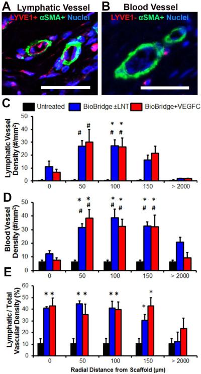
Fig. 3. Immunohistochemistry analysis of lymphatic and blood vascular regeneration.
A–B. Confocal microscopy images of immunofluorescently stained (A) lymphatic collectors (LYVE1+, α-SMA+) and (B) blood-vascular arterioles (LYVE1−, α-SMA+) in the lymphedematous right limb [24]. Scale bar, 50 μm. C–D. Quantitative analysis (#/mm2) of the density of (C) lymphatic collectors and (D) blood-vascular arterioles within regional rings, as defined by indicated radial distances from the border of the scaffold. E. Lymphatic fraction of total (blood + lymphatic) vascular density in percent (n ≥ 3). *p < 0.05 versus untreated irradiated tissue (control group), #p < 0.05 versus surrounding irradiated tissue at > 2000 μm for the same animal, based on ANOVA with Holm's post adjustment.

