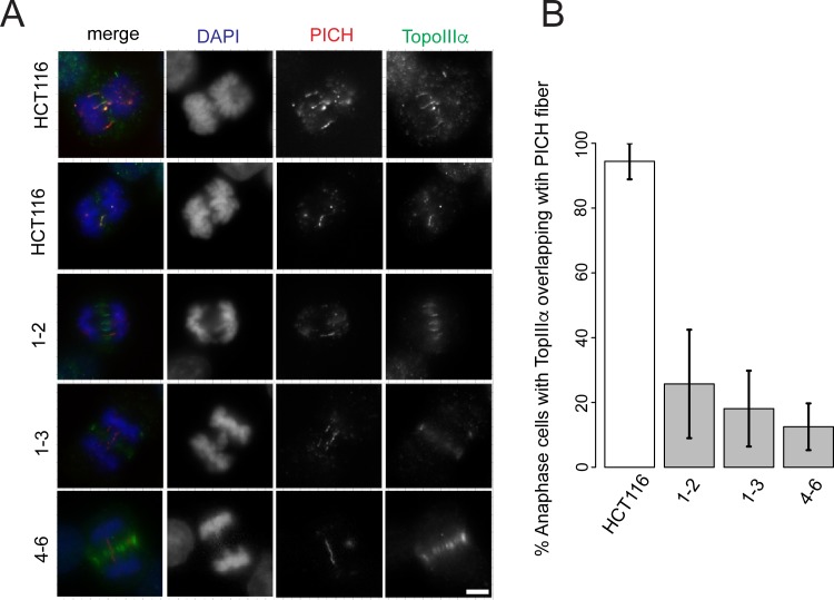
Fig 7. RMI2 is necessary for localization of TopoIIIα to anaphase UFBs.
Representative images of RMI2 wild-type and null HCT-116 cells (A) stained with anti-TopoIIIα (green), anti-PICH (red) and DAPI for DNA (blue). Scale bar 5 μm. (B) Quantification of anaphase B HCT-116 wild-type and RMI2 null cells detected with PICH and TopoIIIα. Numbers represent only the pool of anaphase B cells with PICH fibers and whether these demonstrated colocalization with TopoIIIα. The data was taken from three independent experiments, with a minimum of nine anaphase cells per experiment per cell line.

