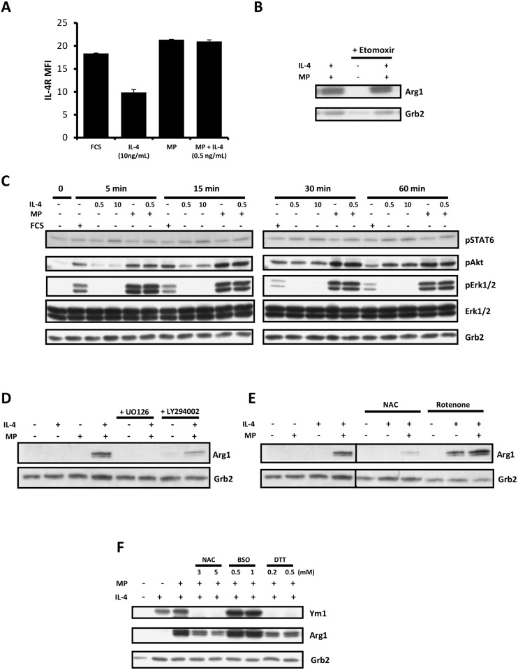
Fig 3. MP enhances IL-4-induced M2 skewing via ROS and activation of Erk and Akt in MФs.
A) Surface IL-4R expression on WT MФs cultured for 48 h ± 10 ng/mL IL-4, 5% MP, or 5% MP + 0.5 ng/mL IL-4, as assessed by FACS using PE-conjugated anti-IL-4Rα. B) Western blot of WT MФs exposed to 5% MP ± etomoxir (100 μM) ± 0.5 ng/mL IL-4 for 72 h and probed for Arg1. Grb2 was used as a loading control. C) Western blot of WT MФs treated or not with 10% FCS, 5% MP ± 0.5 ng/mL or 10 ng/mL IL-4 for 5, 15, 30 or 60 min. The blot was probed for pSTAT6, pAKT, pERK1/2 and ERK1/2. Grb2 was used as a loading control. D) Western blot of WT MФs exposed for 72 h to the indicated combinations of low dose IL-4 (0.5ng/mL) and 5% MP ± the MEK inhibitor U0126 (5 μM) or the PI3K inhibitor LY294002 (5 μM). E) Western blot of WT MФs exposed for 72 h to the indicated combinations of low dose IL-4 (0.5ng/mL) and 5% MP ± N-acetylcysteine (NAC, 2 mM) or rotenone (0.25 μM). F) Western blot of WT MФs exposed for 72 h to the indicated combinations of low dose IL-4 (0.5ng/mL) and 5% MP ± low and high doses of N-acetylcysteine (NAC), L-buthionine-S,R-sulfoximine (BSO), or dithiothreitol (DTT). All results are representative of 2 or 3 independent experiments.

