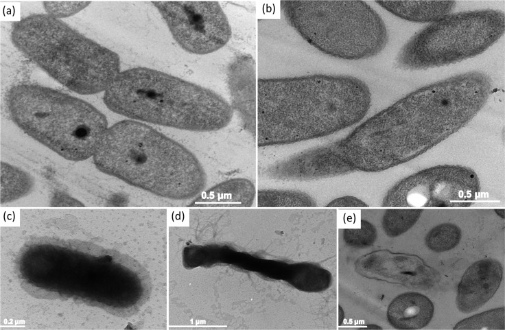
Fig 8. Ultrastructural changes of B. pseudomallei 316c cells after exposure to AgNPs.
TEM micrographs prepared by ultramicrotome for ultra-thin sections show (a) untreated cells, (b-d) cells exposed to 1/5 MIC of AgNPs and (e) cells exposed to MBC for 1 hr at 37°C. Dashed arrows show nucleoid of B. pseudomallei 316c, and asterisk indicates bacterial vacuole.

