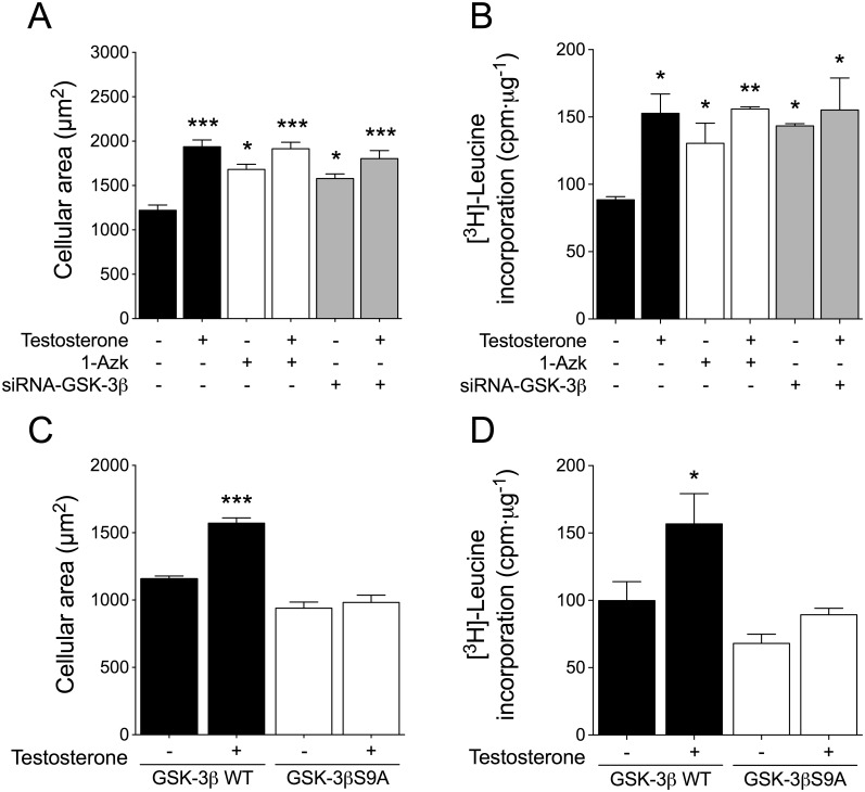
Fig 7. GSK-3β inhibition is involved in testosterone-induced cardiac myocyte hypertrophy.
Cardiac myocytes were pretreated with 1-Azk (1 μM) or transfected with siRNA-GSK-3β (A and B, respectively), or the cells were transfected with GSK-3βWT or GSK-3βS9A expression plasmid (C and D, respectively) and then stimulated with 100 nM testosterone for 48 h. For cell size measurement, cardiac myocytes were incubated with CellTracker Green and examined by confocal microscopy (n > 80 cells from 4 independent cell cultures). Protein synthesis was determined by [3H]-leucine incorporation. The data correspond to the ratio (basal counts/min)·μg-1 protein for each experimental condition (n = 6 for each condition). Values are presented as the mean ± SEM. *p < 0.05, **p < 0.01, and ***p < 0.001 vs. control non-stimulated condition.

