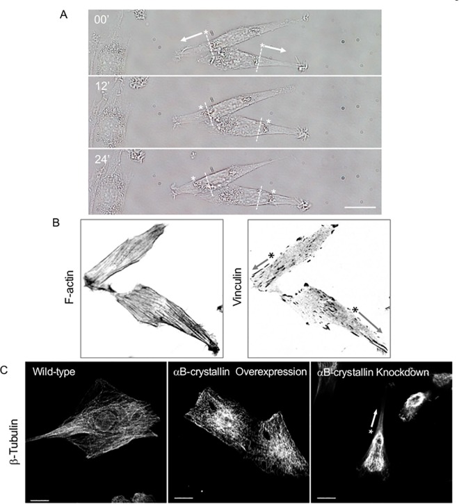
Fig 12. Time-lapse imaging of αB-crystallin knockdown L6 cells.
Directional cell migration as fast as about 10 μm/ h are typically seen. Position of the nuclei are indicated by asterisks and arrows indicate the migratory direction. Bar is 20 μm (A). Asymmetric distribution of nucleus, F-actin stress fiber, and focal adhesion of αB-crystallin knockdown cells (B, inverted contrast image of Fig 11) are coincide with asymmetric polymerization of microtubule caused by αB-crystallin knockdown. Bar is 20 μm (C).

