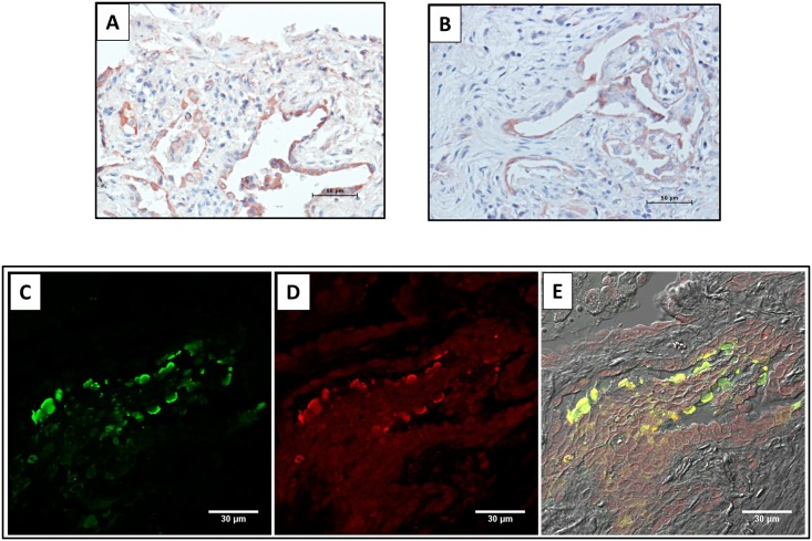
Fig 4. Localization of CC16 in alveolar epithelial cells.
Panels A and B: IPF lung tissues illustrating positive staining for CC16 in pneumocytes. Panels C-E: Immunofluorescence double labeling for CC16 (green) and SP-C (red) demonstrated the colocalization of both proteins in the alveolar epithelium of an IPF lung (yellow).

