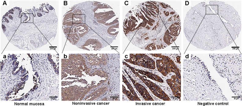
Fig 1. Representative images of Derlin-1 protein expression in IHC microarrays.
(A and a), normal mucosa; (B and b), noninvasive cancer; (C and c), invasive cancer; (D and d), negative control; IHC = immunohistochemistry (A, B, C and D, magnification ×40, scale bars: 200 μm; a, b, c and d, magnification ×200, scale bars: 50 μm).

