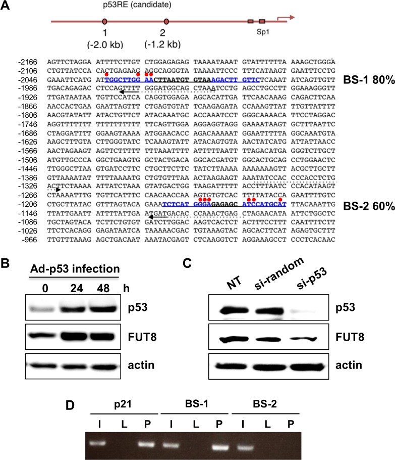
Fig 1. Identification of the p53-DNA binding site within the FUT8 genomic promoter.
(A) Mapping of p53-binding DNA sequences on the FUT8 genomic locus. Based on DNA sequence results, each candidate shows high homology to the p53-DNA binding consensus sequence (as indicated by%). Red points indicate mismatched residues compared with the consensus sequence, whereas candidate sites are written in blue. (B) Protein expression of p53 and FUT8 in HepG2 cells was analyzed by western blot after 24- and 48-h of infection with Ad-p53. (C) FUT8 protein expression after p53 knock-down in HepG2 cells was determined by western blot. (D) ChIP assay of HepG2 cells infected with Ad-p53 and Ad-LacZ. The PCR product for the p21 binding site of p53 was used as a positive control. I: 10% input, L: Ad-LacZ infection, P: Ad-p53 infection. NT: no treatment. Experiments were carried out in triplicate and repeated at least three times.

