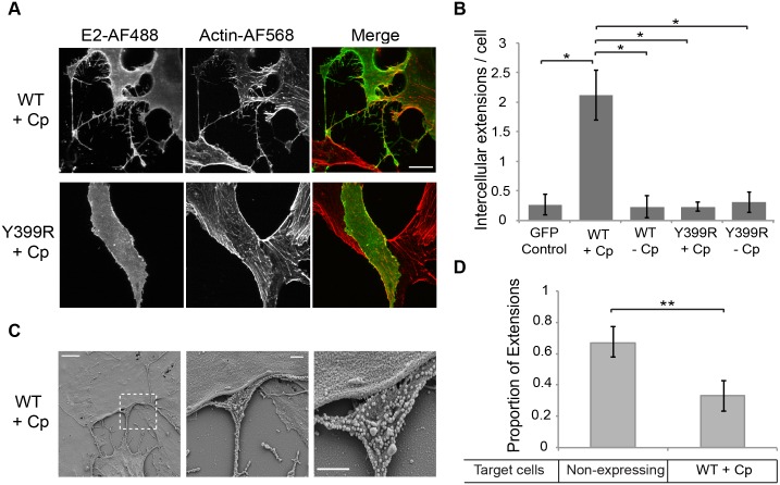
Fig 9. The alphavirus structural proteins induce the formation of intercellular extensions that preferentially target non-expressing cells.
(A) Vero cells were transfected with plasmids encoding the SFV structural proteins with or without capsid and with or without the E2 Y399R mutation. At 24 h post-transfection the cells were fixed, permeabilized, immunostained for the SFV E2 protein and stained with phalloidin-Alexa568 to detect F-actin. Images were acquired by confocal microscopy and are representative of three independent experiments. Images from one optical section are shown. Bar = 20 μm. (B) Using the conditions described in (A), the number of intercellular extensions per E2-expressing cell was quantitated based on positive staining for actin and contact with a neighboring cell. The graph represents the mean and standard deviation of three independent experiments. *P<0.05. (C) Vero cells were transfected with a plasmid encoding the SFV structural proteins including capsid, incubated at 37°C for 24 h, fixed, processed for SEM and imaged using a Zeiss Supra 40 field emission SEM. Shown are representative SEM images. Left panel: Bar = 5 μm. Middle panel: Bar = 1 μm. Right panel: 2X zoom of middle panel, bar = 1μm. (D) Vero cells were transfected with a plasmid encoding the SFV structural proteins including capsid, incubated at 37°C for 24 h, fixed, permeabilized, and immunostained for the SFV E2 protein and α-tubulin. The intercellular extensions per expressing cell were quantitated based on their positive staining for tubulin and their contact with another cell, and scored based on expression of SFV E2 in the target cell. The graph represents the mean and standard deviation of three independent experiments. ** P<0.01.

