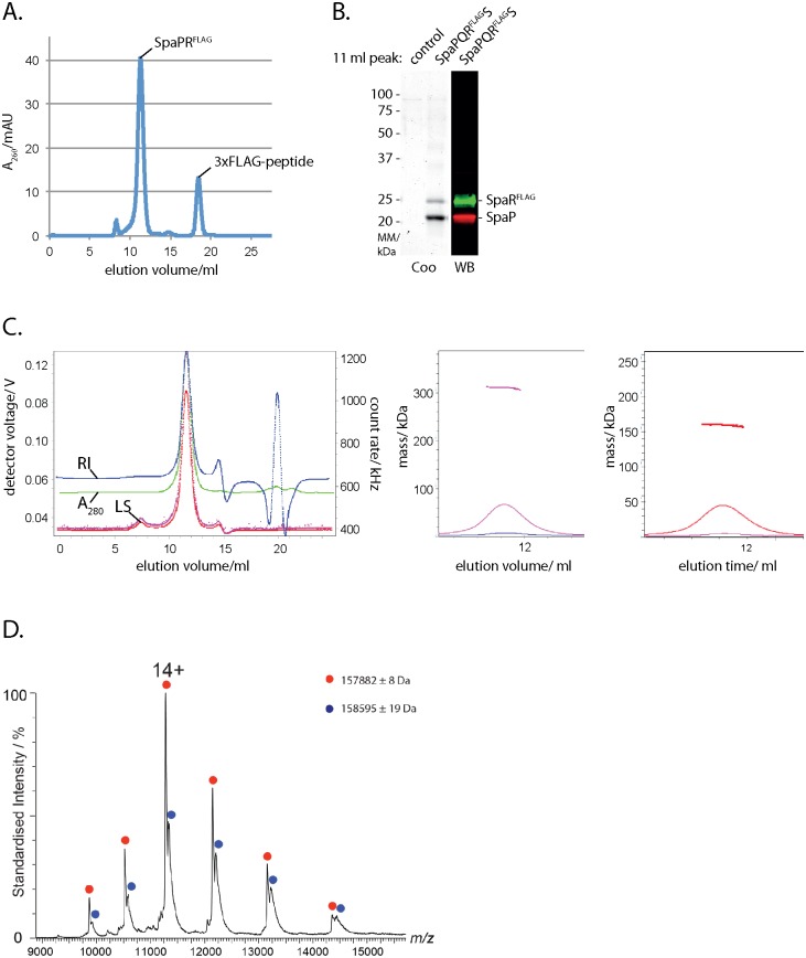
Fig 1. Isolation and stoichiometry analysis of the SpaPR subcomplex of the needle complex.
(A) Elution profile of the purified SpaPRFLAG complex run on a Superdex 200 10/300 GL column. The peaks corresponding to the SpaPRFLAG complex and 3xFLAG peptide are indicated. (B) Coomassie-stained SDS PAGE gel of purified SpaPRFLAG complex and of its FLAG-deficient control (left). Immunodetection of SpaP (green) and SpaRFLAG (red) on Western blot from purified SpaPRFLAG complex separated by SDS PAGE (right). (C) Traces of indicated detector signals from size exclusion chromatography—multi angle laser light scattering of purified SpaPRFLAG complex (left). ASTRA-calculated mass profile of total components of peak of purified SpaPRFLAG complex (polypeptides and detergent, middle). ASTRA-calculated mass profile polypeptide components of peak of purified SpaPRFLAG complex (right). (D) Native mass spectrum of the SpaPRSTREP complex. Peak series corresponding to the SpaP:SpaRSTREP complex in a 5:1 ratio is marked in red, with the most abundant charge state (14+) indicated. The peak series marked in blue corresponds to the same SpaPR complex bound to a ligand with a mass of approximately 710 Da, indicative of an associated phospholipid. Note that the measured mass for SpaPR heterohexamer (157.882 kDa) is heavier than the theoretically calculated mass (157.280 kDa). Abbreviations: Coo: Coomassie stained, WB: Western blot, RI: refractive index, LS: light scattering.

