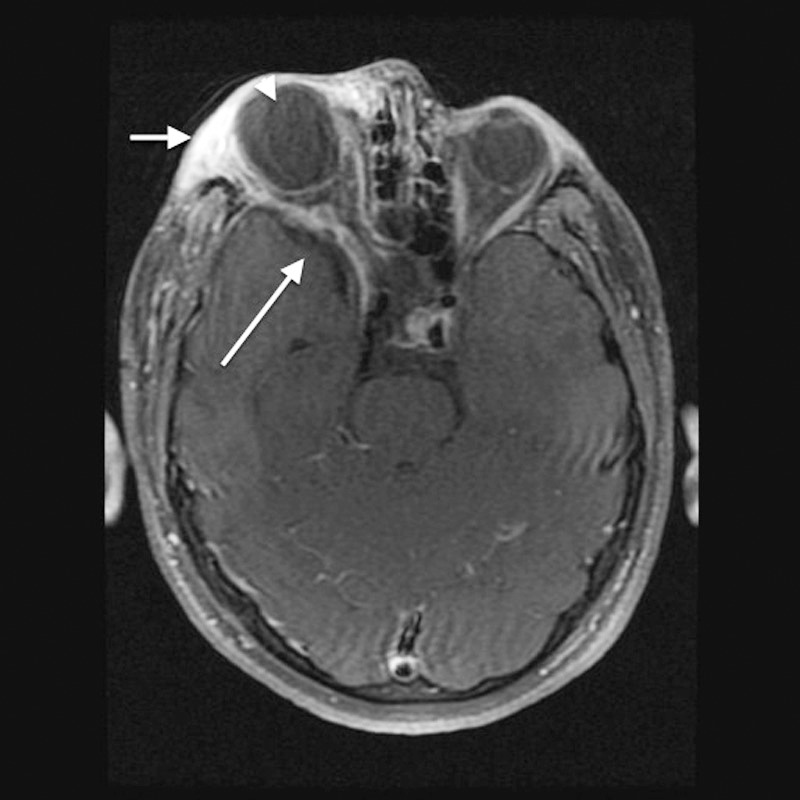
Figure 3.

Axial T1 fat-saturated contrast-enhanced magnetic resonance imaging shows diffuse edema with heterogeneous enhancement of the preseptal soft tissue and superolateral extraconal right orbit consistent with plexiform neurofibroma (short arrow). Enlargement of the right middle cranial fossa as a result of greater sphenoid wing dysplasia is visible (long arrow). The globes are asymmetric in size, with marked buphthalmos on the right (arrowhead).
